Quantitative Wood Anatomy is a field that scientifically studies the variability of structural characteristics of xylem in trees, shrubs, and herbaceous species. Its main objective is to understand the relationships between plant function, development, and environmental conditions. Xylem not only transports water and nutrients, but also stores sugars and hormones and provides mechanical support to the plant. Thus, each xylem structure has its own specific function and is largely fixed at the time of its formation.
Xylem characteristics can often be localized within annual growth rings, allowing scientists to understand how the plant’s structure and function changed in a particular year. Such studies also help us understand how sensitive plants are to environmental changes. However, generating large-scale xylem anatomical data is fraught with several methodological challenges. This involves proper sample collection, microsection preparation, image capturing through microscopy, and image analysis, each step of which can introduce measurement errors ranging from 5 to 30%. If these errors are not corrected, it becomes difficult to draw meaningful conclusions from data with small differences. Therefore, a rigorous protocol and quality control are essential.
Introduction: The Importance of Xylem and Quantitative Studies
Xylem plays a central role in the life processes of plants. It not only transports water and nutrients, but also stores hormones and sugars and provides stability to the leaves, flowers, and the entire canopy. The diverse structures of xylem have evolved in countless ways to perform different functions. Quantitative wood anatomy scientifically studies this diversity. It not only helps understand the relationship between structure and function but also reveals environmental influences at the level of annual growth rings. For example, the differences between earlywood and latewood cells indicate the time of their development and the environmental conditions at that time.
In fact, both internal and external factors influence the development of xylem structure. Sometimes, poorly adapted xylem structure can even lead to tree death. Studies in this field primarily focus on conducting cells such as vessels and tracheids, parenchyma (axial and radial), and fibers.
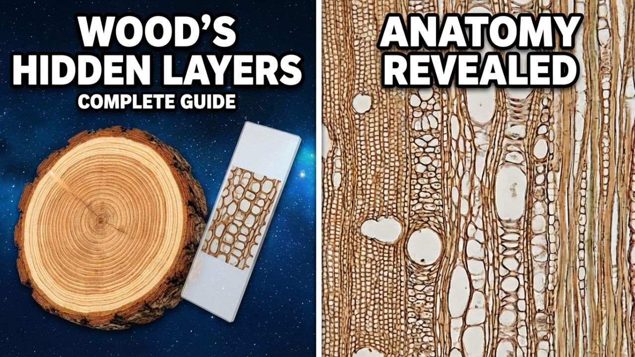
Diversity and Challenges of Xylem Structures
Xylem anatomical features are extremely minute and sensitive. Measuring them requires extreme care and precision. It is also essential that the samples are sufficiently large and representative to ensure reliable results. Higher quality samples, high-resolution images, and the use of modern image analysis tools can yield more accurate data.
Recent improvements in computer performance, automated image analysis systems, and data interpretation techniques have made it possible to collect more quantitative and detailed data than ever before. As a result, quantitative wood anatomy is being used in several research areas, including climate and growth interactions, stress response, tree functionality, and the identification of climate change-resistant species.
Step-by-Step Process: From Sample Collection to Data
Step 1: Sample Collection in the Field
For quantitative wood anatomy, information is obtained from the structures of stems, shoots, branches, roots, rhizomes, and even leaves of monocot and dicot plants. An increment borer is often used for this purpose. A sharp borer and precise drilling in the radial direction are crucial during sample collection. For small trees, herbs, or roots, samples can be taken by digging around the roots using standard gardening tools. Pruners and small-toothed saws are used for branches and small stems. It is essential to remove the first part of the sample to minimize the possibility of cracking or tearing during microsectioning.
Step 2: Preparation of Microsections
General Procedure
Microsections are typically 10–20 microns thick. A sledge or rotary microtome is used for this purpose. Stains such as safranin, Astra Blue, toluidine blue, and cresyl violet are used to color the cell walls and enhance contrast on the slide.
In some cases, it is necessary to boil or simply soak the samples in water, embed them in paraffin, or use a corn starch/rice starch solution. This prevents damage to the cell structures during cutting.
Microtome Blades
The sharpness and quality of the blade are extremely important. Dull blades can damage the delicate structures. It is often best to change the blade after each sample or use a fresh section of the blade. Special blades are required for hard tissues. High-quality blades from companies like Leica and Feather are recommended.Excellent results are obtained.
Step 3: Image Capturing and Analysis
The prepared slides are imaged using an optical microscope. Ensuring high-resolution images and proper contrast is essential. Subsequently, quantitative analysis of structures such as conducting cells, fibers, and parenchyma is performed using specialized software tools.
It is important to note that measurement errors can occur at each stage, ranging from 5–30%. These errors are particularly significant in smaller cells. Therefore, quality control and adherence to rigorous protocols are crucial.
Conclusion: Quality and Scientific Significance
Quantitative wood anatomy is a powerful tool for understanding plant growth, function, and environmental impacts. With properly prepared samples and rigorous data collection protocols, scientifically sound and reliable conclusions can be drawn. This field not only illuminates plant structure and function but also contributes to important research areas such as climate change, stress response, and tree longevity.
Overall, this technique is extremely useful in understanding the microstructures of plants and revealing their functional significance. As image analysis and data processing techniques advance, quantitative wood anatomy is providing researchers with more accurate and detailed data, opening new possibilities in environmental and botanical sciences.